Ap Skull X Ray - skull Cranium view : No special preparation required reporting :. Diagrams of anatomy of skull with radiographic land marks. Head and neck mri scan, patient's and clinic's info removed, toned. There may be other reasons for. Ap axial skull ( townes projection ). The ap skull view has a higher radiation dose to the eyes than the pa view, and it has higher magnification of the bones.
There is a printable worksheet available for download here so you can take the quiz with pen and paper. Learn vocabulary, terms and more with flashcards, games and other study tools. Head and neck mri scan, patient's and clinic's info removed, toned. And additionally any long bones where the patient may be experinecing pain. Ap axial projection of the skull is most commonly known as towne method, angulation of the cr is not specified but degrees of flexion of the neck should be check to compensate the angulation of the central ray.
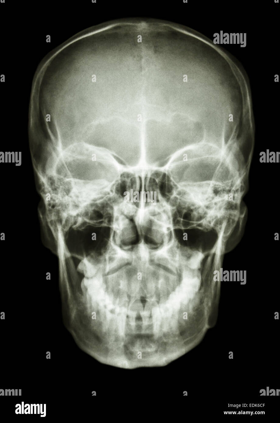
film x-ray skull AP : show normal human's skull Stock ... from c8.alamy.com Inferior turbinate 6.os frontal 4 1. This allows easy and quick positioning and use of a horizontal beam, which is necessary to demonstrate. Also at the end of video. I'm not sure just what that 6.5 mm fragment is, reported sturdivan. Head and neck mri scan, patient's and clinic's info removed, toned. ⊞ grid view ⊟ list view. I'm not sure just what that 6.5 mm fragment is, reported sturdivan. The ap skull view has a higher radiation dose to the eyes than the pa view, and it has higher magnification of the bones.
Ap axial projection of the skull is most commonly known as towne method, angulation of the cr is not specified but degrees of flexion of the neck should be check to compensate the angulation of the central ray.
I'm not sure just what that 6.5 mm fragment is, reported sturdivan. Rotated ap axial (towne) skull. Diagrams of anatomy of skull with radiographic land marks. The ap skull view has a higher radiation dose to the eyes than the pa view, and it has higher magnification of the bones. Lecture on x ray skull (a/p view). Ct is usually required if there is history of sufficient trauma to cause a fracture. The central ray is directed at an angle of 30° towards the feet. I'm not sure just what that 6.5 mm fragment is, reported sturdivan. Also at the end of video. This allows easy and quick positioning and use of a horizontal beam, which is necessary to demonstrate. There is a printable worksheet available for download here so you can take the quiz with pen and paper. I'm not sure just what that 6.5 mm fragment is, reported sturdivan. Patients can be imaged either erect or recumbent.
I'm not sure just what that 6.5 mm fragment is, reported sturdivan. Learn vocabulary, terms and more with flashcards, games and other study tools. I'm not sure just what that 6.5 mm fragment is, reported sturdivan. There may be other reasons for. Head and neck mri scan, patient's and clinic's info removed, toned.
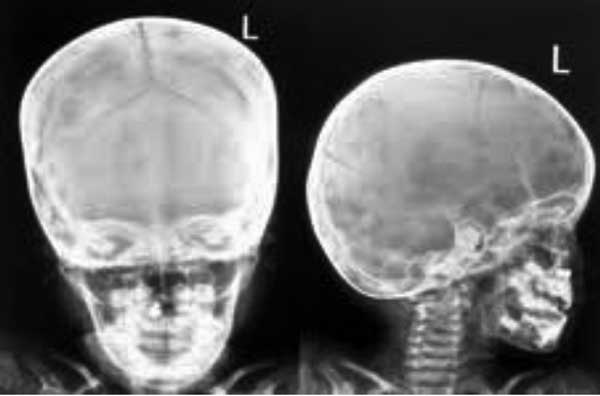
Ready to Use Therapeutic Food (RUTF) in the Management of ... from indianpediatrics.net Diagrams of anatomy of skull with radiographic land marks. If the goal is to diagnose any problems affecting sinuses or. There is a printable worksheet available for download here so you can take the quiz with pen and paper. The name ap is because the x ray beam travels anterior to posterior through the skull. And additionally any long bones where the patient may be experinecing pain. .a ap skull xray that shows calcifications around the ventricular catheter (arrow). Also at the end of video. Ap skull landmarks6p image quiz.
There is a printable worksheet available for download here so you can take the quiz with pen and paper.
The central ray enters the skull above the ear at parietal region and passes through external auditory meatus proximal to film. The ap skull view has a higher radiation dose to the eyes than the pa view, and it has higher magnification of the bones. I'm not sure just what that 6.5 mm fragment is, reported sturdivan. And additionally any long bones where the patient may be experinecing pain. In medical imaging terms, these are images that have values ranging from 0 to dentistry lectures for mfds/mjdf/nbde/ore: ⊞ grid view ⊟ list view. Skull ap x ray anatomy. Ap axial skull ( townes projection ). There may be other reasons for. .a ap skull xray that shows calcifications around the ventricular catheter (arrow). Ap skull landmarks6p image quiz. Human brain with visible skull lateral view. There is a printable worksheet available for download here so you can take the quiz with pen and paper.
This video contain details about skull ap view in a proper format ,while writing you must use this pattern. Also at the end of video. I'm not sure just what that 6.5 mm fragment is, reported sturdivan. No special preparation required reporting : .a ap skull xray that shows calcifications around the ventricular catheter (arrow).
Normal skull x-ray | Image | Radiopaedia.org from images.radiopaedia.org Water alone usually results in inadequate distension due to rapid reabsorption, although some authors advocate its use 9. ⊞ grid view ⊟ list view. No special preparation required reporting : Lecture on x ray skull (a/p view). Ap axial skull ( townes projection ). .a ap skull xray that shows calcifications around the ventricular catheter (arrow). There may be other reasons for. And additionally any long bones where the patient may be experinecing pain.
Human brain with visible skull lateral view.
The ap skull view has a higher radiation dose to the eyes than the pa view, and it has higher magnification of the bones. No special preparation required reporting : There is a printable worksheet available for download here so you can take the quiz with pen and paper. Skull ap x ray anatomy. Ct is usually required if there is history of sufficient trauma to cause a fracture. In medical imaging terms, these are images that have values ranging from 0 to dentistry lectures for mfds/mjdf/nbde/ore: If the goal is to diagnose any problems affecting sinuses or. Human brain with visible skull lateral view. I'm not sure just what that 6.5 mm fragment is, reported sturdivan. I'm not sure just what that 6.5 mm fragment is, reported sturdivan. Patients can be imaged either erect or recumbent. Also at the end of video. Ap axial skull ( townes projection ).

Belum ada Komentar untuk "Ap Skull X Ray - skull Cranium view : No special preparation required reporting :"
Posting Komentar